Research Article | | Peer-Reviewed

Phylogenetic and Pathogenic Characterization of Mauginiella scaettae as the Causal Agent of Date Palm (Phoenix dactylifera L.) Inflorescence Rot in Khozestan & Bushehr Provinces of Iran

Received: 3 May 2025     Accepted: 3 June 2025     Published: 30 June 2025
Views:       Downloads:
Abstract

Inflorescence rot or Khamedj, is a devastating disease of date palm (Phoenix dactylifera L.) in Iran but has not been extensively characterized. Mauginiella scaettae Cav. 1925 (Pleosporales; Ascomycota), causal agent of this disease was isolated from infected male and female inflorescences of date palm from different regions in the Khozestan and Bushehr provinces southwest of Iran. Twelve fungal isolates were collected from infected spathes however, SCUA-Am-133 strain of M. scaettae was used to infect healthy male inflorescence of date palms to satisfy Koch’s postulates but follow on assessments were based on GA. Phylogenetic reconstruction using the rRNA sequence data ITS region found no intraspecific differentiation of strains and confirmed the placement of M. scaettae within the family Phaeosphaeriaceae. Our characterization provides new insights into this inflorescence rot to allow better detection and management of the disease. This is the first report of molecular identification M. scaettae the causal agent of date palm in Khozestan and Bushehr provinces of Iran.

Published in Science Development (Volume 6, Issue 2)
DOI 10.11648/j.scidev.20250602.13
Page(s) 38-42
Creative Commons

This is an Open Access article, distributed under the terms of the Creative Commons Attribution 4.0 International License (http://creativecommons.org/licenses/by/4.0/), which permits unrestricted use, distribution and reproduction in any medium or format, provided the original work is properly cited.

Copyright

Copyright © The Author(s), 2025. Published by Science Publishing Group

Keywords

Date Palm, Inflorescence Rot, Mauginiella Scaettae ITS Region

1. Introduction
Date palm (Phoenix dactyliferaL.) is considered as the "tree of life" as it is one of the most important crops in Iran, where it has been cultivated for thousands of years. Date palm is mostly cultivated for fruit; it is also grown in many countries as an ornamental or landscape tree . This tree is a strategic crop for many countries, including Iran, with a total production of more than 1 307 908 tons annually . Currently, more than 460 date palm cultivars have been cultivated in different areas of Iran, including provinces of Bushehr, Hormozgan, Kerman, Khuzestan, and Sistan & Baluchistan . The date palms are affected by several plant pathogens, including fungi. Pathogenic fungi cause diseases in date palms that not only reduce the quantity and quality of the fruit, but also in some cases lead to the death of the trees and offshoots .
Losses due to pests and pathogens are also significant with serious damage linked to Bayoud disease which infects palm fronds and is caused by Fusarium oxysporum f. sp. albedinis . One of the major diseases of date palm is inflorescence rot (known as Khamedj disease in Iran). Inflorescence rot is a major factor that limits yield, which in severe cases can destroy around 20-60% of the date palm inflorescences. The disease is most devastating during extended periods of heavy rain before the emergence of the spates which are bracts covering the developing flowers. The symptoms are more apparent on the internal face of the spates and infections occur early in the young inflorescences when the spates are still hidden in the leaf bases. This disease can persist on the palms and cause rotted inflorescences for several years. Mauginiella scaettae is frequently isolated infected inflorescences of date palm and, as such, is thought to be the causal agent. Date palm is inflorescence rot was first reported by Carava, (1925) in Cyrenaica, Libya and throughout the region ; Egypt , the Iraq ; and Southern Spain . However, other fungi such as Fusarium oxysporum Schldl. emend. Snyder and Hansen, Fusarium moniliforme Sheld. Aggregate, Fusarium solani (Mart.) Appel and Wollenw. aggregate Snyder and Hansen, Thielaviopsis paradoxa (Dade) C. Moreau. and Trichothecium roseum (Pers.) Link, have also been commonly found associated with rotten date palm inflorescences . This study aimed to identify the fungi that are associated with date palm inflorescence rot in Khozestan and Bushehr that M. scaettae, isolated from rotten tissues of Phoenix dactylifera inflorescences, is introduced using both morphological and molecular phylogenetic approaches.
2. Materials and Methods
2.1. Sample Collection and Isolation of Fungal Strains
In 2021, to identify the fungal associated with inflorescences rot of date palm, 12 symptomatic samples of infected spathes and inflorescences of date palms with necrosis and rotted symptoms were collected from different areas in the regions of Abadan, AbPakhsh, Bushehr, Behbahan, Khorramshahr, Susangerd, Shadegan, Shabankareh, Ramhormoz, Mahshahr and Hoveyzeh in Bushehr and Khuzestan provinces, in the southwest of Iran. The samples were packed in plastic bags and brought to the laboratory on ice. The isolation procedure was done according to Abass (2013) . The plant fragments were sectioned into small pieces (approx.1–2 cm) and surface-sterilized by dipping them in a 5% solution of sodium hypochlorite for 5 min, followed by rinsing three times in distilled water and drying on sterile filter paper. The plant pieces were placed on potato dextrose agar (PDA; Merck, KGaA) in Petri dish plates and incubated at 25 °C for 5-7 days. The individual colonies were sub-cultured on fresh PDA plates and purified by a single-spore isolation method, as described by Abdullah et al. (2005) . Among them, in this study, 12 morphologically-identical isolates of M. scaettae, fungi were obtained, which subjected to morphological and molecular identification. For micro-morphological observations, mycelial samples were obtained from cultures grown on PDA and imaged using a Nikon Coolpix digital camera installed on an Olympus BX50 light microscope. The description of the mycelium and arthroconidia follows established terminologies .
2.2. Pathogenicity Test
Arthroconidia from SCUA-Am-133 strain were grown on PDA were used for pathogenicity assays, and with a suspension of arthroconidia (106 spores.m l-1) prepared in sterile water. Healthy (male) spates were surface-sterilized with 95% ethanol, and the upper external surface of the spate was removed so that the flowers and strands were exposed. These were inoculated by spraying with 20 mL of the arthroconidia suspension per inflorescence, placed in commercially obtained sterile paper bags and incubated at condition in the garden. Spathes were examined for symptoms after 4-7 days.
3. Morphological and Physiological Study
The morphological observations and culture characteristics of the fungal isolates were determined by sub-culturing them on PDA media and incubating at 25 °C under 12 h light-dark cycles for 7-14 days. Slide cultures were prepared according to the protocol described by Beneke & Rogers (1996) . Sporulation structures, including conidia, conidiophores and conidiogenous cells, were measured using 100x objective lens of a Leitz Wetzlar (SM-LUX) Basic Biological light microscope. The Measurements for each fungal structure (at least fifty) were reported as a maximum and minimum range in association with 95% confidence limits and standard deviation. The photomicrographs were taken using an OLYMPUS BX-50 light microscope fixed with a TUCSEN GT 12 digital camera.
4. DNA Extraction and PCR Amplification
A single-spore culture of each isolate was grown on PDA at 2°C under a dark condition for 7–14 days to achieve the maximum mycelial growth. The fungal biomass of the agar surface was collected using a sterile glass slide and powdered in the mortar containing liquid nitrogen using a pestle. Genomic DNA of each isolate was extracted from the powdered mycelia using a phenol- and chloroform-based organic method . DNA samples were analyzed using a spectrophotometer (eppendorf Bio Photometer plus) and loading on the agarose gel. Partial regions of ITS, were amplified using primer pairs ITS1/ITS4 . Respectively, Polymerase chain reaction (PCR) mixture contained 5 μl of 10x prime Taq Reaction Buffer (GenBio, South Korea), 3 μl of MgCl2 (25 mM), 1.5 μl of dNTPs (2.5 mM each), 1.5 μl of each primer (10 μM), 0.6 μL of Prime Taq DNA Polymerase (5 units/μl, GenetBio, South Korea), and 2 μl of template DNA (125 ng/μL) adjusted with purified water (Mili-Q Water) to a final volume of 50 μl. Amplification reactions were performed in a thermal cycler (MJ MiniTM Gradient Thermal Cycler) and started with an initial denaturation step at 95°C for 3 min, followed by 35 cycles of 94°C for 30 s, 56°C (ITS), 58°C for 40 s and 72°C for 1 min, followed by a final elongation step at 72°C for 5 min. The quality of PCR products was checked by agarose gel electrophoresis in 1x Tris-acetic acid-EDTA (TAE) buffer.
5. Purification and Sequencing
The PCR products were size-fractionated by electrophoresis in 1% agarose gel (Hispan Agar) immersed in 1.0×Trisacetic acid-EDTA (TAE) buffer stained with commercial safe stain (Sina Colon, Iran) and checked for smearing and target/non-target DNA bands. Pure products were directly sent to NiaGen Company (Tehran, Iran) for sequencing using forward and reverse primers. For non-pure products, DNA bands of the expected size were excised and purified by GF-1 Ambi Clean Kit (Vivantis, Malaysia) according to the manufacturer’s instructions. The Bio Edit Sequence Alignment Editor Version 7.0.9.0 and DNA Baser Sequence Assembler v4 programs (2013, Heracle BioSoft, www. Dna Baser. com) were used to edit and assembly forward and reverse sequences, respectively. Newly generated sequences were deposited in GenBank.
6. Phylogenetic Analyses
Sequence management was conducted within the Geneious Prime bioinformatics package . Sequences were aligned using MAFFT using default setting and trimmed to remove flanking 18S/28S sequences. Phylogenetic reconstruction was conducted using PhyML (Maximum Likelihood) and the GTR (Generalised time reversible) substitution model with 1000 bootstrap replications .
7. Results
7.1. Date Palm Inflorescence Rot Symptom
The disease is generally first observed when spots appear in late winter and early spring. Infected spathes of male first exhibit rot symptoms when they begin to emerge in early spring. Typically, these are observed as necrotic spots and patches on the spathe. When the spathe matures and splits open, white mycelia is observed covering the flowers, especially at the top of the spathe. Hyphae are visible on the outside of the spathe. Female inflorescences, the flowers erupt rapidly from the spathe. Necrotic and rot symptoms are seen from flower emergence in late spring onwards, as pollination is in progress. Generally, symptoms begin as a white mycelial mass attacking the flowers. The spathes split to reveal partial to near complete coverage of the flowers with white mycelium. As the infected flowers emerge, the areas previously covered with white mycelium become necrotic, killing the mature flowers (Figure 1 a-c).
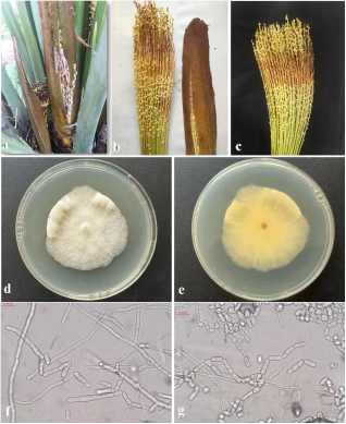
Figure 1. a-c: symptoms of necrosis and rotting of inflorescences, d and e: 7-day SCUA-Am-133 colony of M. scaettae in PDA culture from the upper and lower surface, f and g: round and cylindrical conidiophores, conidia and arthroconidia.
7.2. Morphological characteristics of M. Scaettae
The fungal colonies on PDA grew slowly and their diameter reached 4.7 cm after seven days at 25°C. The colonies of the isolates studied occupied the entire surface of the Petri dish for three weeks. The colony margins on the culture medium were usually smooth; however, in some isolates they were wavy. The color of the upper surface of the colony on PDA medium was generally creamy white and on the lower surface of the Petri dish, it was creamy yellow to pale lemon. The growth of hyphae in the Petri dish on the surface of the PDA medium was fluffy and in some cases, were formed aerial, thick, cottony and dense hyphae (Figure 1 d-e).
The hyphae were colorless, transparent, highly branched, 7.5-0.8 μm in diameter and an average diameter of 3.5 μm. Abundant arthrospores were produced by fragmentation and bundling of aerial hyphae. Arthrospores were unicellular or multicellular (1-6 cells and rarely up to 17 cells), most arthrospores were unicellular or bicellular, colorless, cylindrical or elliptical and rarely round (Figure 1 f-g). The size range of round unicellular conidia was usually 7.5-20 μm and the size range of cylindrical conidia (mostly 1-4 cells) was unicellular 7.5×37-7.5×10, bicellular 7.5×35-7.5×20, tricellular 7.5×47-7.5×22, quadricellular 5×50-42×10.5 μm. The separation of conidia in this fungus occurs in a schizolytic manner at the transverse dividing wall. The cylindrical conidia have a rounded, flat end or, in a number of conidia, conical and pointed, which is sharp at the point of separation of the conidia from each other. Rattan & Al-Dboon (1980) believe that this fungus, in addition to producing arthrospore-type conidia, can form a series of other conidia on the conidiophore. In this study, the formation of such conidia on short conidiophores was observed in some cases.
7.3. Pathogenicity of M. Scaettae
The pathogenicity test of M. scaettae on the spathes of the Barhi cultivar was carried out by isolate SCUA-Am-133 in the palm grove of the Date and Tropical Fruit Research Institute (Figure 3). For this purpose, three healthy spathes were selected and a slit was made on their surface with a sterile scalpel. Then, discs from the growing edge of the seven-day fungal colony on the PDA culture were removed with a cork borer and placed in the slit made on the surface of the spathe. After inoculation, the split part of the spathe was covered with Para film. Then, to prevent the spikelets from spreading and to facilitate and accurately assess the percentage of disease severity, the tip of the spot and the spikelets were closed with plastic tape. PDA medium without fungus was also used for the control treatment. Symptoms of the disease appeared 7 days after inoculation.
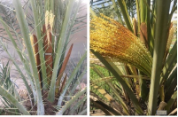
Figure 2. Symptoms and effects of Mauginiella scaettae isolate SCUA-Am-133 on inflorescences of Barhi cultivar, A: control and B: treatment.
7.4. Phylogeny of Mauginiella scaettae Cavara, (1925)
The results of the BLAST search of the studied isolate showed that ITS sequence has 100 nucleotide similarity with the type strain M. scattae CBS 23958. In the single-gene phylogenetic tree based on the ITS region sequence for Mauginiella isolates (Figure 3), this isolate clustered with the type strain M. scattae CBS 23958 and created an evolutionary branch with a bootstrap value of 100%.
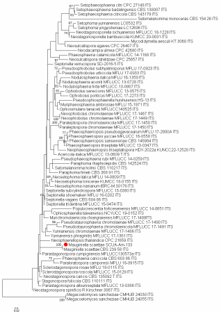
Figure 3. Phylogenetic tree of isolates belonging to the M. scaettae obtained in maximum likelihood analysis based on ITS sequences (the examined sample is shown with a red solid circle).
8. Discussion
Inflorescence rot is one of the most devastating diseases of date palm. M. scaettae were isolated from the infected spathes of male and female date palm, it was suggested to be the causal agent. However, in our study M. scaettae was the only isolated from male and female date palm inflorescences. Symptoms of the disease were observed on all parts of the inflorescences, which includes brownish or rusty areas visible on the non-opened spathe after the pathogen has already invaded the floral tissues. It is agreement with previous reports . After 10 days of growth on PDA at 22°C only white fungal colonies were observed, composed of branched hyaline septate hyphae. This is consistent with published descriptions of M. scaettae, whose mycelia and arthroconidia suggested it to be an anamorph of an unknown ascomycete .
Abbreviations

GA

Geographic Atrophy

PDA

Potato Dextrose Agar

ITS

Internal Transcribed Spacer

PCR

Polymerase Chain Reaction

TAE

Trisacetic Acid-Ethylenediaminetetraacetic Acid

MAFFT

Multiple Alignment Using Fast Fourier Transform

GTR

Generalised Time Reversible

Conflicts of Interest
The authors declare no conflicts of interest.
References
[1] Abass, M. H. 2013. Microbial contaminants of date palm (Phoenix dactylifera L.) in Iraqi tissue culture laboratories. Emir. J. Food Agric. 25, 875–882.
[2] Abdullah, S. K., Asensio, L., Monfort, E., Gomez-VidalS., Palma-Guerrero, J. and Salinas, J. 2005. Occurrence in Elx, SE Spain of inflorescence rot disease of date palms caused by Mauginiella scaettae. Journal Phytopathology, 153(7-8): 417–22.
[3] Amani, M., Farokhinejad, R., & Mehrabi-Koushki, M. 2023. Xenoacremonium palmarum sp. nov., a novel species associated with Phoenix dactylifera in Iran. Phytotaxa, 632(2): 165-174.
[4] Beneke, E. S. and Rogers, A. L. 1996. Medical mycology and human mycoses. Star Publishing Company, Belmont, 239 pp.
[5] Bensaci, M. B., Toumatia, O., Bouras, N., Rahmania, F., Douglas, B., Wade, S., Griffith, G. W., Luis A. J. 2023. Phylogenetic and pathogenic characterization of Mauginiella scaettae as the causal agent of date palm (Phoenix dactylifera L.) inflorescence rot in southeast of Algeria. Physiological and Molecular Plant Pathology 127, 102062.
[6] Bouguedoura, N., Bennaceur, M., Babahani, S., & Benziouche, S. E. 2015. Date palm status and perspective in Algeria. Chapter 4 in Book: JM Al-Khayri et al.(eds.), Date Palm Genetic Resources and Utilization. Africa and the Americas.
[7] Cavara, F. 1925. Mauginiella scaettae Cav. nuovo ifomicete parassita della palma da datteri in virenacia, Bollettino del Real Orto Botanico di Napoli 8: 207–211.
[8] Chabrolin, C. 1928. La pourriture de inflorescence du palmierdattier. Ann Epiphyt, 14, 377-414.
[9] Chabrolin, C. (1930). Les maladies du Dattier. Journal d'agriculture traditionnelle et de botanique appliquée, 10(107), 557-566.
[10] Drummond, A. J., Ashton, B., Buxton, S., Cheung, M., Cooper, A., Duran, C., Field, M., Heled, J., Kearse, M., Markowitz, S., Moir, R., Stones-Havas, S., Sturrock, S., Thierer, T., Wilson, A. 2011. Geneious. v5.4 Geneious v5.4.
[11] El-Modafar, C. 2010. Mechanisms of date palm resistance to Bayoud disease: Current state of knowledge and research prospects. Physiological and Molecular Plant Pathology 74(5-6): 287-294.
[12] FAO. 2021 Food and Agriculture Organization of the United Nations.
[13] Guindon, S., Dufayard, J. F., Lefort, V., Anisimova, M., Hordijk, W., Gascuel, O. 2010. New algorithms and methods to estimate maximum-likelihood phylogenies: assessing the performance of PhyML30, Syst. Biol. 59(3) 307–321.
[14] Hameed, M. A. 2012. Inflorescence rot disease of date palm caused by Fusarium proliferatum in Southern Iraq, Afr. J. Biotechnol. 11(35) 8616–8621,
[15] Hall, T. A. 1999. BioEdit: a user-friendly biological sequence alignment editor and analysis program for windows 95/98/NT. In Nucleic Acids Symposium Series, 41: 95–98.
[16] Hussain, F. 1958. Occurrence of date palm inflorescence rot in Iraq, Plant Dis. Rep. 42, 1958.
[17] Katoh, K., Misawa, K., Kuma, K. I., & Miyata, T. 2002. MAFFT: a novel method for rapid multiple sequence alignment based on fast Fourier transform. Nucleic acids research, 30(14), 3059-3066.
[18] Mehrabi-Koushki, M., Khodadadi-Pourarpanahi, S., & Jounbozorgi, S. (2018). Fungal endophytes associated with some thermotolerant plants in salt-stress ecosystem. Mikologiya i Fitopatologiya, 52(3), 187-195. Микология и фитопатология 52(3): 187-195.
[19] Michael, I. F., Sabet, K. A. 1970. Biology and control of Mauginiella scaettae Cav, the pathogen of Khamedj disease in the united Arab Republic, Annual Date Grower’s Institue 47: 5–8.) 555.
[20] Rattan, S. S. and Al-Dboon, A. H. H. 1981. Notes on fungi associated with date palm I. Sydowia, 33: 246-264.
[21] White, T. J., Bruns, T., Lee, S. J. W. T., & Taylor, J. 1990. Amplification and direct sequencing of fungal ribosomal RNA genes for phylogenetics. PCR protocols: a guide to methods and applications, 18(1), 315-322.
Cite This Article
  • APA Style

    Amani, M., Zad, R. F., Mehrabi-Koushki, M. (2025). Phylogenetic and Pathogenic Characterization of Mauginiella scaettae as the Causal Agent of Date Palm (Phoenix dactylifera L.) Inflorescence Rot in Khozestan & Bushehr Provinces of Iran. Science Development, 6(2), 38-42. https://doi.org/10.11648/j.scidev.20250602.13

    Copy | Download

    ACS Style

    Amani, M.; Zad, R. F.; Mehrabi-Koushki, M. Phylogenetic and Pathogenic Characterization of Mauginiella scaettae as the Causal Agent of Date Palm (Phoenix dactylifera L.) Inflorescence Rot in Khozestan & Bushehr Provinces of Iran. Sci. Dev. 2025, 6(2), 38-42. doi: 10.11648/j.scidev.20250602.13

    Copy | Download

    AMA Style

    Amani M, Zad RF, Mehrabi-Koushki M. Phylogenetic and Pathogenic Characterization of Mauginiella scaettae as the Causal Agent of Date Palm (Phoenix dactylifera L.) Inflorescence Rot in Khozestan & Bushehr Provinces of Iran. Sci Dev. 2025;6(2):38-42. doi: 10.11648/j.scidev.20250602.13

    Copy | Download

  • @article{10.11648/j.scidev.20250602.13,
      author = {Majid Amani and Reza Farokhyne Zad and Mehdi Mehrabi-Koushki},
      title = {Phylogenetic and Pathogenic Characterization of Mauginiella scaettae as the Causal Agent of Date Palm (Phoenix dactylifera L.) Inflorescence Rot in Khozestan & Bushehr Provinces of Iran
    },
      journal = {Science Development},
      volume = {6},
      number = {2},
      pages = {38-42},
      doi = {10.11648/j.scidev.20250602.13},
      url = {https://doi.org/10.11648/j.scidev.20250602.13},
      eprint = {https://article.sciencepublishinggroup.com/pdf/10.11648.j.scidev.20250602.13},
      abstract = {Inflorescence rot or Khamedj, is a devastating disease of date palm (Phoenix dactylifera L.) in Iran but has not been extensively characterized. Mauginiella scaettae Cav. 1925 (Pleosporales; Ascomycota), causal agent of this disease was isolated from infected male and female inflorescences of date palm from different regions in the Khozestan and Bushehr provinces southwest of Iran. Twelve fungal isolates were collected from infected spathes however, SCUA-Am-133 strain of M. scaettae was used to infect healthy male inflorescence of date palms to satisfy Koch’s postulates but follow on assessments were based on GA. Phylogenetic reconstruction using the rRNA sequence data ITS region found no intraspecific differentiation of strains and confirmed the placement of M. scaettae within the family Phaeosphaeriaceae. Our characterization provides new insights into this inflorescence rot to allow better detection and management of the disease. This is the first report of molecular identification M. scaettae the causal agent of date palm in Khozestan and Bushehr provinces of Iran.
    },
     year = {2025}
    }
    

    Copy | Download

  • TY  - JOUR
    T1  - Phylogenetic and Pathogenic Characterization of Mauginiella scaettae as the Causal Agent of Date Palm (Phoenix dactylifera L.) Inflorescence Rot in Khozestan & Bushehr Provinces of Iran
    
    AU  - Majid Amani
    AU  - Reza Farokhyne Zad
    AU  - Mehdi Mehrabi-Koushki
    Y1  - 2025/06/30
    PY  - 2025
    N1  - https://doi.org/10.11648/j.scidev.20250602.13
    DO  - 10.11648/j.scidev.20250602.13
    T2  - Science Development
    JF  - Science Development
    JO  - Science Development
    SP  - 38
    EP  - 42
    PB  - Science Publishing Group
    SN  - 2994-7154
    UR  - https://doi.org/10.11648/j.scidev.20250602.13
    AB  - Inflorescence rot or Khamedj, is a devastating disease of date palm (Phoenix dactylifera L.) in Iran but has not been extensively characterized. Mauginiella scaettae Cav. 1925 (Pleosporales; Ascomycota), causal agent of this disease was isolated from infected male and female inflorescences of date palm from different regions in the Khozestan and Bushehr provinces southwest of Iran. Twelve fungal isolates were collected from infected spathes however, SCUA-Am-133 strain of M. scaettae was used to infect healthy male inflorescence of date palms to satisfy Koch’s postulates but follow on assessments were based on GA. Phylogenetic reconstruction using the rRNA sequence data ITS region found no intraspecific differentiation of strains and confirmed the placement of M. scaettae within the family Phaeosphaeriaceae. Our characterization provides new insights into this inflorescence rot to allow better detection and management of the disease. This is the first report of molecular identification M. scaettae the causal agent of date palm in Khozestan and Bushehr provinces of Iran.
    
    VL  - 6
    IS  - 2
    ER  - 

    Copy | Download

Author Information
  • Tropical Fruits Research Center, Shaeed Chamran University, Ahwaz, Iran

  • Shaeed Chamran University, Ahwaz, Iran

  • Shaeed Chamran University, Ahwaz, Iran